Expert Consensus on the Clinical Application of Bronchoscopy in Secondary pneumonitis After Solid Tumor Treatment
DOI:
https://doi.org/10.65364/iomj.2026.04Abstract
Patients with solid tumors who have undergone anti-tumor therapy are at high risk of developing pneumonia. The etiologies include both infectious and non-infectious factors (e.g., immune-related pneumonia, drug-induced pulmonary toxicity, and radiation pneumonitis). Given that etiological diagnosis can be challenging in some cases, bronchoscopy is required to facilitate differential diagnosis and guide subsequent clinical management. Currently, there is a lack of specific clinical guidelines addressing this issue. Under the auspices of the the China Respiratory Oncology Collaboration (CROC) Zhejiang Collaboration Group, and based on the internationally accepted Delphi method, an expert consensus on bronchoscopy for newly developed pneumonia in solid tumor patients post anti-tumor therapy was formulated following multiple rounds of meetings, discussions, and revisions. This consensus covers key aspects including the timing, indications, contraindications, pre-procedural preparation, selection of bronchoscopic techniques, specimen testing requirements, and interpretation of bronchoscopic findings. It is anticipated that this consensus will help standardize the practice of bronchoscopy for newly developed pneumonia in solid tumor patients after anti-tumor therapy, and improve the diagnostic efficacy and procedural safety.
BACKGROUND
Cancer patients are a high-risk population for pneumonia due to a confluence of factors, including underlying comorbidities, the immunosuppressive and/or immune dysregulation effects of anticancer therapies, and other iatrogenic causes. The etiology of pneumonia in this population is complex and can be broadly categorized into infectious and non-infectious causes. Infectious agents comprise bacteria, fungi, viruses, and parasites. A meta-analysis investigating lung infection pathogens in Chinese lung cancer patients (mostly post-chemotherapy) revealed that Gram-negative bacteria predominated (63%), led by Klebsiella pneumoniae (17%), Pseudomonas aeruginosa (14%), and Escherichia coli (13%)[1]. Conversely, non-infectious pneumonitis is a direct consequence of anticancer treatments themselves. This includes conditions like immune checkpoint inhibitor pneumonitis (CIP), which has a reported incidence of potentially 5%-19% in real-world settings, and radiation pneumonitis, a common complication of thoracic radiotherapy with incidence of 16%-36%[2-16]. Interstitial lung disease associated with small-molecule tyrosine kinase inhibitors (TKIs) - including agents targeting epidermal growth factor receptor (EGFR), anaplastic lymphoma kinase (ALK), cellular-mesenchymal epithelial transition factor (c-MET), rearranged during transfection (RET), ROS proto-oncogene 1 (ROS1), breakpoint cluster region-Abelson (BCR-ABL), and v-raf murine sarcoma viral oncogene homolog B (BRAF) - occurs with a reported incidence of 0.4% to 5.3%[17-20]. Thus, the overall incidence of secondary pulmonary inflammation in cancer patients is considerable. Furthermore, the common coexistence of infectious and non-infectious etiologies introduces notable diagnostic dilemmas[21]. As therapeutic approaches for infectious and non-infectious pneumonia differ entirely, misdiagnosis or delayed intervention is associated with severe implications for patients’ prognosis.
The clinical and radiological presentation of non-infectious pneumonitis in cancer patients - including symptoms such as cough, dyspnea, and fever, and imaging findings like ground-glass opacities and consolidation - is often non-specific and overlaps considerably with that of pulmonary infections or tumor progression[2,11,22-24]. In this context, bronchoscopy has become an increasingly critical tool for etiological differentiation. Studies have demonstrated its utility in identifying characteristic inflammatory patterns. For instance, in patients with CIP, bronchoalveolar lavage (BAL) reveals lymphocytosis and inverted CD4/CD8 ratio[22,25,26]. Furthermore, transbronchial lung biopsy can provide histopathological evidence to clarify the diagnosis[25,27]. Therefore, a comprehensive diagnostic approach integrating serology, imaging, bronchoscopy, and pathology is often essential to establish a timely and accurate diagnosis, guide appropriate therapy, and prevent clinical deterioration due to misdiagnosis.
However, as an invasive procedure, bronchoscopy carries risks of complications -including bleeding, hypoxemia, arrhythmia, and pneumothorax - that pose additional threats to this vulnerable population[28-30]. Consequently, a meticulous pre-procedural evaluation by a specialist is mandatory. This assessment must rigorously examine respiratory and vital organ function, baseline oncologic status, and the degree of immunosuppression to strictly determine indications and contraindications.
Current domestic and international bronchoscopy guidelines are largely designed for general or immunocompetent populations and fail to address the distinct clinical challenges in cancer patients. This population frequently presents with unique risk factors - such as coagulopathy, compromised cardiopulmonary reserve, and neutropenia - that significantly elevate the procedural risk. This critical gap in existing guidelines underscores the clinical necessity of the present expert consensus. This consensus provides a systematic approach to bronchoscopy in cancer patients with pneumonitis following anticancer therapy. It aims to standardize procedures, guide the selection of diagnostic projects and laboratory tests, facilitate accurate interpretation of results, assist in personalized risk-benefit assessment, and enhance safety protocols, thereby optimizing diagnostic accuracy and therapeutic outcomes.
SCOPE OF APPLICATION
This consensus applies to patients with various solid tumors who present with new-onset pulmonary infiltrates following anticancer therapy (e.g., chemotherapy, targeted therapy, radiotherapy, or immunotherapy). These infiltrates, indicative of parenchymal or interstitial inflammation, may stem from diverse infectious or non-infectious causes, including treatment-related conditions such as radiation, drug-induced, or CIP.
The document specifically addresses clinical scenarios requiring bronchoscopy for etiological clarification or treatment guidance. Covered procedures include conventional bronchoscopy, BAL, protected specimen brush (PSB), bronchial mucosal biopsy, transbronchial lung biopsy (TBLB), transbronchial cryobiopsy (TBCB), endobronchial ultrasound-guided transbronchial needle aspiration (EBUS-TBNA), and rigid bronchoscopy.
METHODS
Expert panel composition
This consensus was jointly initiated by the China. Respiratory Oncology Collaboration (CROC) Zhejiang Collaboration Group. Experts were organized to extensively solicit opinions from a multidisciplinary panel, including those from pulmonology and critical care medicine, radiology, radiation oncology, clinical laboratory, pathology, infectious diseases, and general thoracic surgery, based on current domestic and international evidence-based data and clinical experience. The final version was reviewed and finalized by the core expert group.
Literature search
A comprehensive literature review Literature searches were conducted in the following Chinese and English databases: Chinese databases: China Biomedical Literature Database (CBM), Wanfang Data, and China National Knowledge Infrastructure (CNKI); English databases: PubMed, Embase and and ClinicalTrials.gov databases.
Formation of the consensus
The consensus statements were formulated following the internationally accepted Delphi method. First, an expert panel for consensus development was established. After conducting literature searches, the drafting group developed the consensus framework. Following multiple rounds of deliberations and expert voting, 18 recommendation statements were finalized and the final level of expert consensus was specified for each statement. The expert voting options were categorized into three levels: Agree, Neutral, and Disagree. Consensus on a given item was defined as achieved when the proportion of experts voting Agree exceeded 70%.
PRE-PROCEDURAL EVALUATION, DECISION-MAKING AND TIMING
Question 1: Timing of bronchoscopy
Recommendation 1: Early bronchoscopy is recommended in the absence of contraindications - specifically, when there is no improvement after 48-72 h of empirical treatment (e.g., persistent fever, no alleviation of clinical symptoms) or upon disease progression (e.g., worsening hypoxemia, expansion of radiographic lesions) - to establish an etiological or pathological diagnosis and guide subsequent treatment. (Consensus level 100%)
In the diagnostic evaluation of secondary pulmonary inflammation in patients with solid tumors following anticancer therapy, hematological and imaging studies have certain limitations in determining the underlying cause. Although bronchoscopy can provide critical diagnostic information, it is an invasive procedure with inherent risks and potential complications. Therefore, its timing must be carefully considered. A balance must be struck between the patient’s clinical status and procedural risks to avoid both premature intervention without clear indication and delayed examination when it is critically needed.
It is recommended that bronchoscopy be performed early in cases of non-response to 48-72 h of empirical treatment or upon disease progression, in order to identify pathogens or obtain pathological confirmation to guide further management[31]. If the patient’s condition deteriorates rapidly to acute respiratory distress syndrome (ARDS), or in the presence of high-risk factors such as active massive hemoptysis or uncontrolled acute myocardial infarction, a multidisciplinary team (MDT) discussion should be initiated to thoroughly evaluate the benefit-risk profile of the procedure and determine the optimal timing for intervention.
MDT
Question 2: Clinical departments constituting the MDT assessment
Recommendation 1: For patients with multiple comorbidities or critically ill patients at high risk of bronchoscopy, and in whom the etiology of pulmonary inflammation remains unclear - requiring differentiation among causes such as infection, drug-induced lung injury, immune-related pneumonitis, and tumor progression - a multidisciplinary discussion is recommended prior to performing bronchoscopy. (Consensus level 90%)
An MDT is fundamental to ensuring the safe performance of bronchoscopy. This team should include physicians from relevant specialties such as Respiratory and Critical Care Medicine, Medical Oncology, Infectious Diseases, Radiology, Rheumatology and Immunology, Microbiology, and Pharmacology, and Anesthesiology. If the patient has specific comorbidities, corresponding departments should be involved in the decision-making process. For example, patients with coronary heart disease (especially those with a history of coronary stent implantation) require evaluation by a cardiologist; those with cerebrovascular disease necessitate input from a neurologist. The MDT should conduct a comprehensive assessment focusing on both the “necessity” and “feasibility” of bronchoscopy.
Question 3: Components of MDT assessment
Recommendation 1: The MDT discussion covers topics such as the patient’s baseline condition and organ function, the status of tumor treatment, the severity of pneumonia, the necessity of bronchoscopy, and the specific content of the examination. (Consensus level 98%)
(1) Patient’s baseline condition and organ function risk.
a. Performance status: This assesses the patient's ability to perform daily activities and their overall health condition. An ECOG score of 4 indicates a high-risk factor for procedures, necessitating careful evaluation of the procedure’s necessity and associated risks[32].
b. Control of comorbidities: This includes management of acute or subacute conditions such as hypertensive crises, diabetic ketoacidosis or hyperosmolar states, acute cardiovascular events, aortic dissection, pulmonary embolism, and severe electrolyte imbalances. Any uncontrolled acute conditions should be prioritized for correction. If stabilization proves difficult, a thorough risk-benefit analysis for the procedure should be conducted.
c. Respiratory and circulatory system function: This refers to patients with Type I or Type II respiratory failure (especially those with PaO2/FiO2 ≤ 200 mmHg, or decompensated respiratory acidosis), severe airway stenosis or obstruction, decompensated heart failure, malignant arrhythmias, or circulatory instability such as shock[33,34]. These patients are at high risk for bronchoscopy and should be approached with extreme caution.
(2) Disease-related factors evaluation.
a. Tumor-related factors: This includes the type and stage of the tumor, as well as an estimation of the patient’s survival based on tumor biology and clinical progression. For patients with a short-expected survival (e.g., < 3 months), the benefit of invasive procedures should be carefully reconsidered.
b. Treatment-related factors: A comprehensive inquiry into the patient’s recent history of anti-tumor treatments and the treatment phase is required (whether they are in the post-chemotherapy myelosuppression period). It is essential to clarify their use of chemotherapy, targeted therapy, radiotherapy, and immunotherapy. Additionally, the potential risk of treatment-related pulmonary damage should be carefully considered, such as targeted therapy-related pneumonia, CIP, or radiation-induced pneumonitis.
c. Pneumonia severity, necessity of bronchoscopy, and examination content: Tools like the CURB-65 score should be used to objectively assess pneumonia severity. A score of ≥ 2 indicates a higher risk for bronchoscopy, requiring careful evaluation of the procedure's benefits and risks. Imaging assessments should be performed to evaluate pulmonary involvement and lesion characteristics. The necessity of bronchoscopy in identifying the etiology of pneumonia and guiding treatment. Furthermore, the specific type of bronchoscopy and required tests for the patient should be discussed.
Question 4: Indications for bronchoscopy
(1) Suspected infectious pneumonia failing empirical anti-infective therapy, requiring lower respiratory tract specimen acquisition for pathogen identification;
(2) Suspected infection with specific pathogens (e.g., Aspergillus, Mucorales, Nocardia, Mycobacterium tuberculosis, nontuberculous mycobacteria, Pneumocystis jirovecii, viruses, etc.), requiring precise specimen sampling for targeted testing (e.g., metagenomic next-generation sequencing);
(3) Suspected non-infectious pneumonitis (e.g., CIP, radiation pneumonitis, drug-induced pneumonitis, lymphangitic carcinomatosis, etc.) failing empirical treatment, requiring lower respiratory tract specimens to exclude infection or tissue biopsy for differentiation;
(4) Conditions such as obstructive pneumonia secondary to airway obstruction, retention of copious purulent sputum, tracheoesophageal fistula, tracheomediastinal fistula, or bronchopleural fistula, requiring bronchoscopic local interventions (e.g., drainage of purulent secretions, airway stenting).
Question 5: Contraindications to bronchoscopy
(1) Acute coronary syndrome (ACS)
Bronchoscopy is generally not recommended in patients with acute myocardial infarction (within 1 month of onset) or unstable angina (uncontrolled). Routine bronchoscopy should be performed 4-6 weeks after ACS, following evaluation by a cardiologist[35]. However, ACS should not be considered an absolute contraindication when clinical assessment indicates potential benefits outweigh the risks[36].
(2) Active massive hemoptysis
Bronchoscopy carries a high risk during active massive hemoptysis, as the procedure may exacerbate bleeding and induce asphyxia. If bronchoscopy is deemed essential, preparations for establishing an artificial airway and emergency resuscitation must be in place[37,38].
(3) High risk of bleeding
Bronchoscopy is generally not recommended in cases of uncorrected coagulopathy (INR > 1.4), platelet count (PLT) < 20 × 109/L, or active bleeding tendency (e.g., uncontrolled hemophilia)[35,39]. Transbronchial lung biopsy should be considered relatively contraindicated when PLT is < 60 × 109/L[39]. Bronchoscopy may be considered after transfusion to improve platelet count and coagulation function.
(4) Vascular abnormalities at risk of rupture
Bronchoscopy carries a significant risk in patients with thoracic aortic aneurysm, aortic dissection, or esophageal varices, due to the potential for procedure-induced stress or trauma to precipitate rupture.
(5) Other high-risk conditions
In patients with conditions such as malignant arrhythmia, severe cardiopulmonary insufficiency, hypertensive crisis, severe pulmonary hypertension, increased intracranial pressure, acute cerebrovascular event, severe psychiatric disease, or extreme systemic debilitation, the risk of complications from bronchoscopy is elevated. The procedure should only be performed after assessment of the risk-benefit ratio by relevant specialists and with full resuscitation preparedness.
PRE-PROCEDURAL PREPARATION AND MANAGEMENT
Management of coagulation and bleeding
Based on the MDT assessment, a personalized preparation plan should be developed for high-risk patients to ensure their safety.
Question 6: Preparations for high-risk airway hemorrhage
Recommendation 1: For patients suspected of having malignancies involving the central airways, major vessels, or large cavitary lesions, a contrast-enhanced chest CT or bronchial artery CTA is recommended prior to bronchoscopy to assess bleeding risk. If the imaging suggests a high risk and bronchoscopy is still indicated, full preparedness for managing massive hemorrhage must be established. (Consensus level 100%)
Recommendation 2: For patients undergoing anti-angiogenesis therapy, if only BAL or PSB is planned, and the patient’s general condition is clinically stable with no coagulopathy, it is considered unnecessary to discontinue the medication in advance. However, if invasive procedures such as biopsies or interventional treatments are planned, it is recommended to stop the medication based on its half-life, unless the procedure is urgent. (Consensus level 100%)
Recommendation 3: In the absence of underlying coagulation disorders or related comorbidities, routine blood and coagulation tests performed within one week prior to bronchoscopy are considered acceptable. For patients with known hematologic diseases or coagulation abnormalities, a more stringent timeframe of 24 h is recommended for obtaining these laboratory indicators to ensure an accurate pre-procedural bleeding risk assessment. (Consensus level 97%)
Post-chemotherapy patients are prone to thrombocytopenia, coagulation abnormalities, or malignant tumors invading the trachea, bronchi, pulmonary arteries, or bronchial arteries, which can lead to an increased risk of airway hemorrhage. Therefore, a comprehensive assessment of the risk of airway hemorrhage is required before the procedure. This includes a detailed inquiry into the patient’s history of bleeding, as well as their medication history, particularly the use of antiplatelet agents, anticoagulants, and anti-vascular-targeted therapies. There is currently no universally established standard for the optimal timeframe for pre-bronchoscopy coagulation tests. Our expert panel reached the following consensus based on clinical practice and patient risk stratification: For patients without underlying coagulation disorders or related comorbidities, routine blood tests and coagulation function tests obtained within one week prior to the procedure are considered acceptable. For patients with known hematologic diseases or coagulation abnormalities, we recommend obtaining these laboratory indicators within 24 h before bronchoscopy to ensure accurate assessment of bleeding risk. Abnormal blood counts or coagulation functions should be corrected proactively. Antiplatelet or anticoagulant medications should be discontinued according to their half-life[34,39,40-44].
In patients with suspected tumor invasion of the central airways, major vessels, or large cavitary lesions, pre-procedural imaging with contrast-enhanced chest CT or bronchial artery CTA is strongly recommended. This evaluation is critical for identifying high-risk features, including pulmonary artery involvement, pseudoaneurysms, or signs of abnormal vasculature such as enlarged/tortuous bronchial arteries or bronchial artery-pulmonary artery shunts. For these high-risk cases, a multi-disciplinary consultation involving specialists from pulmonology, critical care, radiology, thoracic surgery, and anesthesiology is essential to weigh the necessity of bronchoscopy and formulate a comprehensive management plan. Furthermore, emergency protocols for massive hemorrhage must be established, including readiness for endobronchial balloon tamponade, bronchial or pulmonary artery embolization, and emergency thoracotomy[39,45,46].
Anti-angiogenic drugs (e.g., bevacizumab, anlotinib, recombinant human endostatin) inhibit neovascularization and can increase vascular fragility, thereby elevating the risk of bleeding during invasive procedures[47-49]. The decision to withhold these drugs before bronchoscopy should be stratified by procedural risk: (1) Low-risk procedures (e.g., BAL, PSB, airway inspection without biopsy): Drug discontinuation is generally unnecessary in clinically stable patients without coagulopathy. (2) High-risk procedures (e.g., biopsy, interventional therapy): It is recommended to withhold the drug prior to the procedure based on its half-life, unless the situation is urgent. The recommended discontinuation times, based on the pharmacokinetics of these agents, are as follows: bevacizumab for 4-6 weeks, anlotinib for 1 week, and recombinant human endostatin for 24 h. These are general guidelines, and the final decisions must be individualized, incorporating the patient’s complete medication history, comorbidities, and hepatic/renal function.
Infection prevention and support
Question 7: Should prophylactic antibiotics be routinely used to reduce procedure-related infection risk in patients with granulocytopenia or lymphopenia secondary to anticancer treatment?
Recommendation 1: Most patients receiving standard chemotherapy for solid tumors are at low risk and do not require routine prophylactic antibiotics before bronchoscopy. Prophylactic antibiotics are recommended for high-risk patients. (Consensus level 90%)
The probability of secondary bacterial infection following bronchoscopy is approximately 6%-8%, with common pathogens including coagulase-positive or negative staphylococci, non-hemolytic or β-hemolytic streptococci, and Klebsiella species[50,51]. The prophylactic use of antibiotics for bronchoscopy remains controversial in clinical practice, particularly in patients with tumor-related immunosuppression. Multiple studies have clearly shown that routine prophylactic antibiotics after diagnostic bronchoscopy have not been demonstrated to reduce the incidence of post-procedure fever, pneumonia, or endocarditis[52,53]. Therefore, guidelines from the British Thoracic Society (BTS) and others generally do not recommend routine use[54]. However, recent large-scale evidence suggests potential benefits in specific high-risk populations. For example, a nationwide Japanese database study (involving more than 68,000 patients undergoing diagnostic bronchoscopy for noninfectious diseases) demonstrated that oral prophylactic antibiotics (such as aminopenicillins or fluoroquinolones) were significantly associated with a reduction in post-procedure infections (OR 0.60; 95% CI 0.45-0.80), particularly in elderly patients (> 70 years), those with malignancy, or those undergoing biopsy or bronchoalveolar lavage[55]. Nevertheless, this benefit must be weighed against risks such as antimicrobial resistance and the lack of clear improvement in mortality.
Neutropenia (granulocytopenia) and lymphopenia have different mechanisms of immunodeficiency, leading to distinct infection risks and evidence bases. For patients with malignancies who develop neutropenia following chemotherapy, they are classified into low-risk and high-risk groups based on the severity of potential complications. Low-risk patients are those expected to have neutropenia (absolute neutrophil count [ANC] < 0.5 × 10⁹/L) lasting ≤ 7 days, with no or few comorbid conditions; high-risk patients are those with neutropenia lasting > 7 days and/or significant medical comorbidities[56]. Currently, most patients receiving standard chemotherapy for solid tumors are classified as low-risk and do not require prophylactic antibiotics before bronchoscopy. However, for high-risk patients, prophylactic antibiotics are recommended.
Lymphopenia is common in patients receiving anti-tumor therapy or those with hematologic diseases. Radiotherapy, chemotherapy (e.g., cyclophosphamide, cisplatin, methotrexate, and taxanes), targeted therapy (e.g., mTOR inhibitors, tyrosine kinase inhibitors targeting VEGFR or PDGFR), and immune checkpoint inhibitors can induce lymphopenia[57-60]. In a prospective Danish population-based study of 98,344 individuals, lymphopenia (defined as a lymphocyte count below the population’s 2.5th percentile, i.e., < 1.1 × 10⁹/L) was linked to a higher risk of both infection-related hospitalization and mortality[61]. Furthermore, early lymphopenia has also been identified as a risk factor for chemotherapy-induced febrile neutropenia[62] and early death following chemotherapy[63]. Given that evidence regarding antibiotic use during bronchoscopy in lymphopenic patients is limited, the expert panel recommends the prophylactic administration of antibiotics for those with lymphopenia (defined as a lymphocyte count below the population’s 2.5th percentile) undergoing this procedure.
Respiratory and circulatory support
Question 8: Respiratory support preparation, risk stratification, and site selection
Bronchoscopy inherently carries certain respiratory-related risks, and such risks are significantly heightened in cancer patients who have undergone anti-tumor therapy - characterized by potential multi-organ dysfunction and complicated by pneumonitis. Therefore, it is of crucial clinical importance to optimize preoperative risk stratification assessment, formulate a comprehensive respiratory support plan, and select an appropriate procedure site.
Recommendation 1: Pre-procedural risk stratification should be conducted based on the patient’s baseline cardiopulmonary function, type and severity of comorbidities, clinical symptoms, coagulation function parameters, and the complexity of the intended procedure. The level of respiratory support must then be tailored accordingly, commensurate with the assessed risk. (Consensus level 97%)
(1) Patients classified as low-risk (basically normal pulmonary and cardiac function; resting respiratory rate of 12-20 breaths/min; oxygenation index (PaO2/FiO2) ≥ 300 mmHg; platelet count ≥ 100 × 109/L; normal coagulation function; requirement for diagnostic procedures only; and absence of conditions such as airway stenosis or a history of massive hemoptysis) may undergo the procedure in a standard bronchoscopy room, accompanied by oxygen inhalation via nasal cannula and ECG monitoring.
(2) Patients classified as moderate-risk (moderate pulmonary dysfunction; mild cardiac insufficiency (NYHA Class Ⅰ-Ⅱ); resting respiratory rate of 21-25 breaths/min; PaO2/FiO2 of 200-300 mmHg; platelet count of 60-100 × 109/L or mild coagulation abnormalities; requirement for small-scale therapeutic procedures; or presence of mild airway stenosis) should undergo the procedure in a bronchoscopy room equipped with high-flow nasal cannula oxygen therapy (HFNC) or non-invasive positive pressure ventilation (NIPPV). Local epinephrine infusion and a hemostatic balloon should be prepared and readily available.
(3) Patients classified as high-risk (severe pulmonary dysfunction; severe cardiac insufficiency (NYHA Class Ⅲ-Ⅳ); complicated by serious underlying diseases such as uremia, liver cirrhosis, or cerebrovascular disorders; resting respiratory rate > 25 breaths/min; PaO2/FiO2 < 200 mmHg; platelet count < 60 × 109/L; INR > 1.5; requirement for large-scale therapeutic procedures; or presence of severe airway stenosis or a recent history of massive hemoptysis) should undergo the procedure in an ICU or a hybrid operating room, equipped with comprehensive respiratory and circulatory support, such as Cone-Beam Computed Tomography (CBCT). Pre-procedural preparations should include HFNC and NIPPV, with intubation and mechanical ventilation conducted in advance. If necessary, intravenous access for rapid blood transfusion, a complete array of emergency medications, and a hemostatic balloon should be immediately available.
TECHNICAL STANDARDS
Operator qualifications: The procedure should be performed by a highly experienced and senior respiratory endoscopist.
Question 9: How to choose different bronchoscopy techniques?
Bronchoscopic procedures include BAL, PSB, mucosal biopsy, TBLB, TBCB, EBUS-TBNA, and rigid bronchoscopy [Table 1]. The choice of procedure must be based on the patient’s clinical presentation, preliminary diagnosis, therapeutic objectives, and procedural risks. An MDT discussion is recommended when necessary.
| Clinical scenario | Primary diagnostic goal | Recommended bronchoscopic technique | Specimens for analysis |
| Confirmed Infectious, Pathogen Unknown | Rapid and accurate identification of pathogens | 1. Standard white light bronchoscopy (WLB)2. BAL and PSB preferred for diffuse lesions3. BAL or PSB under radial-EBUS/fluoroscopic guidance for focal lesions4. Biopsy is not mandatory (consider bronchial mucosal biopsy, TBLB, or TBNA in specific cases) | 1. Microbiology: Smears and cultures2. mNGS/tNGS3. Pathogen-specific PCR (e.g., for TB, CMV)4. Antigen testing: e.g., Cryptococcal antigen, GM test5. BALF cell count: Elevated neutrophils support acute infection |
| Uncertain: Infectious vs. Non-Infectious | To differentiate between infectious and non-infectious causes | 1. Standard WLB2. Probe-based Confocal Laser Endomicroscopy (pCLE)3. BAL4. TBLB, TBNA, or TBCB in selected cases | 1. BALF: - Microbiology (as in Scenario 1) - Cytology: Lymphocyte subset analysis, cell differential count2. Biopsy (bronchial/TBLB): - ROSE - Histology (H&E, immunohistochemistry) - Special stains (e.g., AFB, GMS, India ink, Masson’s trichrome) - Microbial culture or tissue mNGS |
| Confirmed Non-Infectious, Poor Treatment Response | To confirm specific non-infectious etiology or identify overlap syndromes | 1. Standard WLB2. BAL3. TBLB4. TBCB (if lesions are diffuse and diagnosis remains elusive, after risk-benefit assessment) | 1. Histopathology: - Tissue for H&E, special stains, immunohistochemistry2. BALF Analysis: - Cytology: e.g., Lymphocyte subset analysis - Optional pathogen testing (culture/mNGS): To rule out a missed/overlooked infection |
| Possible Co-existing Infectious and Non-Infectious | To obtain evidence for both infectious and non-infectious causes simultaneously | 1. Standard WLB2. Combined use of BAL and TBLB3. Guided techniques (e.g., radial-EBUS, fluoroscopy) based on lesion location4. TBNA or TBCB in selected cases | Same as Scenario 2 |
Recommendation 1: BAL and PSB are the preferred methods for identifying pathogens in infectious pneumonia and differentiating between infectious and non-infectious pneumonia. It is recommended that all patients undergoing bronchoscopy undergo BAL and PSB. (Consensus level 100%)
Recommendation 2: Airway mucosal biopsy and TBLB are generally not mandatory procedures and are applicable in cases where pathological tissue samples are required for differential diagnosis, treatment guidance, or prognosis assessment. (Consensus level 100%)
Recommendation 3: TBCB is not routinely recommended. It is primarily indicated when the etiology remains unclear after a series of examinations, including clinical presentation, laboratory tests, imaging features, BAL, and conventional TBLB, and when there is a high clinical suspicion of diffuse interstitial lung disease or peripheral pulmonary nodules requiring histological confirmation. (Consensus level 100%)
Recommendation 4: Rigid bronchoscopy is not a conventional diagnostic tool but rather a key technique for managing high-risk complex situations and severe complications. (Consensus level 97%)
BAL
BAL is a procedure in which saline is instilled into the bronchial alveoli via a bronchoscope and then aspirated for the collection of alveolar surface liquid and/or the clearance of material occupying the alveoli. For patients without respiratory failure, a total lavage volume of 100-300 mL is recommended, divided into three to five aliquots. The minimal total volume retrieved should be ≥ 5% of the instilled volume; an optimal return is ≥ 30%. A minimal volume of 5 mL of the pooled BAL sample is required for cellular analysis (optimal 10-20 mL), striking a balance between minimizing lavage volume and ensuring an adequate specimen in patients with respiratory failure[64]. BAL is the preferred method for identifying pathogens in infectious pneumonia and for differentiating between infectious and non-infectious pneumonias. Laboratory analysis of the fluid should be strategic and directed at the relevant differential diagnoses. Tests employed may include ay include cell differential and count, microbiological smears and cultures, fungal antigen testing, pathogen nucleic acid testing, metagenomic next-generation sequencing (mNGS) for lower respiratory tract pathogens, special stains, and cytopathological examination[65-67].
PSB
PSB is a sampling technique that employs a sheathed brush. This brush is advanced through the bronchoscope to the bronchial segment exhibiting the most significant radiographic infiltration, most rapid progression, or endoscopic evidence of purulent secretions to collect lower respiratory tract specimens. Typically performed alongside BAL, PSB is primarily used for pathogen identification in infectious pneumonia and for cytopathological examination. While it samples a smaller area and offers a narrower spectrum of detectable analyses than BAL, its principal advantage lies in obtaining uncontaminated lower respiratory tract specimens via the protected sheath, thereby facilitating reliable microbiological culture and cytopathological studies[68-70].
Bronchial mucosal biopsy
Bronchial mucosal biopsy involves obtaining tissue samples from endobronchial lesions. It is indicated for establishing an etiological diagnosis in cases of bronchoscopically identified mucosal abnormalities, such as nodules, erosions, swelling, or congestion. Notably, antineoplastic agents like immune checkpoint inhibitors and RAF/MEK inhibitors can induce sarcoid-like reactions[71,72]. Hence, this procedure can yield diagnostic value in patients with suspected new-onset sarcoidosis, even in the absence of bronchoscopically visible mucosal changes. In confirmed cases of infectious pneumonia, bronchial mucosal biopsy is generally not necessary. However, it may be considered if infections such as mycobacterium tuberculosis or fungi are suspected, particularly when BAL or PSB results are negative and significant mucosal abnormalities are present[73,74].
Endoscopic imaging techniques
Autofluorescence bronchoscopy, narrow-band imaging bronchoscopy, and confocal laser endomicroscopy (CLE) share the primary design objective of detecting airway tumors and/or pre-cancerous lesions. Each technique, however, provides a distinct view of mucosal pathology - through differences in fluorescence signals, vascular patterns, and microscopic cellular architecture, respectively. Consequently, they offer an irreplaceable advantage over traditional white-light bronchoscopy in the accurate differentiation among inflammation, interstitial lung disease, and neoplasia[75-77].
TBLB
TBLB obtains small lung tissue specimens, providing crucial evidence for pathological diagnosis. It is primarily indicated for lesions located distal to the segmental bronchi that are not visible by conventional bronchoscopy. The TBLB procedure is generally considered to be safe. However, compared to techniques like BAL, PSB, and bronchial mucosal biopsy, TBLB carries higher risks of bleeding and pneumothorax[78]. Not all patients require TBLB, it should be actively considered when the risk assessment is acceptable in the following scenarios: (1) For undiagnosed conditions: When the etiology remains unclear despite clinical presentation, laboratory tests, imaging features, and BAL results; (2) For complex or multifactorial etiologies: Such as suspected drug-associated pneumonia with concurrent infection; (3) When pathological typing is needed to guide treatment decisions; (4) Upon initial treatment failure: For example, when clinically suspected immune drug-related pneumonia shows poor response to empirical corticosteroid therapy; (5) For atypical or focal radiographic presentations: TBLB demonstrates high success rates and diagnostic value for lesions located within the reach of endobronchial ultrasound.
Technically, TBLB includes traditional fluoroscopy-guided approaches and modern guided bronchoscopy techniques. The latter significantly improves the diagnostic yield for peripheral pulmonary lesions, with an average diagnostic sensitivity of approximately 70%, superior to traditional TBLB techniques (approximately 55%)[79,80]. Commonly used guided bronchoscopy techniques currently include radial endobronchial ultrasound (R-EBUS), guide sheath (GS), virtual/electromagnetic navigation, multimodal augmented reality navigation bronchoscopy, cone-beam CT, and robotic bronchoscopy[81]. Among these, navigation bronchoscopy combined with EBUS-GS is the most frequently used in clinical practice.
Cryobiopsy
Cryobiopsy (CB) is a sampling technique that utilizes cryoadhesion to obtain tissue specimens. It comprises two distinct procedures: Endobronchial cryobiopsy (EBCB) for visible lesions and TBCB for peripheral lesions beyond endoscopic visualization. Compared to forceps biopsy (FB), CB is characterized by larger specimen size, superior architectural preservation, and a higher diagnostic yield. For endobronchial lesions, EBCB carries an increased risk of mild-to-moderate bleeding compared to FB[82,83]; it is therefore recommended when FB sampling is inadequate or when a specific pathology necessitates a larger sample. Similarly, TBCB is not recommended for routine use. Its primary indication arises when the etiology remains elusive despite comprehensive evaluation-including clinical, laboratory, radiological, BAL, and conventional TBLB findings-and when there is a high clinical suspicion of a diffuse interstitial lung disease or peripheral pulmonary nodule requiring histological confirmation.
EBUS-TBNA
EBUS-TBNA is a minimally invasive technique that enables real-time ultrasound-guided precise puncture and biopsy of mediastinal and hilar lesions adjacent to the trachea or bronchi, characterized by accurate positioning, operational safety, and high diagnostic efficiency[84,85]. While not routinely recommended, it should be actively utilized in specific scenarios: (1) Etiological differentiation, when imaging reveals extrapulmonary lesions adjacent to airways requiring distinction between treatment-related pneumonia, tumor recurrence, or lymph node metastasis; (2) Diagnosis of specific pathogen infections, such as obtaining lymph node tissue for pathogenic testing in suspected tuberculosis; (3) Evaluation of systemic diseases, particularly when granulomatous diseases like immunotherapy-induced sarcoidosis are suspected, where demonstrating non-caseating granulomas in lymph nodes provides crucial diagnostic evidence.
Rigid bronchoscopy
The rigid bronchoscope provides a safe, reliable, and powerful operative channel, serving as a key technical platform-rather than a routine diagnostic tool-for managing high-risk and complex scenarios[86,87]. Its primary indications include: (1) performing tissue biopsy in high-risk patients when pathological confirmation is essential yet unattainable by conventional diagnostics, and when factors such as high bleeding risk or severe hypoxemia requiring ventilatory support necessitate a secured approach to manage potential complications like major hemorrhage; (2) managing severe treatment-related airway complications, such as those following radiotherapy or immunotherapy, which may involve central airway obstruction from necrotic material or pseudomembranes, or rapidly progressive stenosis and granulomatous proliferation, all requiring urgent intervention under rigid bronchoscopy-including debridement, resection, balloon dilation, ablation, or stent placement.
Question 10: What tests should specimens obtained via bronchoscopy be sent for?
Bronchoscopy-derived specimens are crucial for establishing a definitive diagnosis. For complex pulmonary inflammation secondary to solid tumor therapy, these specimens should be evaluated comprehensively, precisely, and hierarchically with three key objectives: to differentiate between infectious and non-infectious causes; to identify the specific pathogen if infectious; and to determine the precise etiology if non-infectious.
Recommendation 1: Given its high cost, technical complexity, and challenging interpretation, mNGS is reserved for immunocompromised, critically ill, or rapidly deteriorating patients with suspected pulmonary infections, especially when conventional tests are negative. (Consensus level 100%)
Recommendation 2: When targeted Next-Generation Sequencing (tNGS) is negative yet rare or novel pathogens beyond its detection scope are still suspected, mNGS should be considered. (Consensus level 100%)
Recommendation 3: Due to the greater complexity and risks associated with their procurement, tissue specimens should not be the primary specimen for mNGS testing. They may, however, be considered as an alternative or adjunct to bronchoalveolar lavage fluid (BALF). (Consensus level 100%)
Recommendation 4: Histopathological examination is not mandatory for the diagnosis of most infectious or non-infectious pneumonia. Therefore, comprehensive evaluation should integrate clinical, laboratory, and imaging findings. (Consensus level 100%)
BALF and PSB specimens
(1) Microbiological examination
a. Bacterial Smear and Culture: Used for diagnosing bacterial pneumonia, covering both aerobic and anaerobic bacteria.
b. Fungal smear and culture: Aids in detecting fungal infections, including yeasts, molds, and dimorphic fungi.
c. Tuberculosis (TB) smear and culture: Employs acid-fast staining and mycobacterial culture to identify Mycobacterium tuberculosis and non-tuberculous mycobacterial infections. Weakly acid-fast positivity is a characteristic feature of Nocardia infection.
d. Polymerase chain reaction (PCR) based molecular testing: Enables confirmation of infections such as viruses, Legionella, and Pneumocystis jirovecii, and also detects Mycobacterium tuberculosis as well as rifampin resistance through rpoB gene mutation analysis.
e. Antigen testing: Includes assays for Cryptococcus neoformans capsular antigen and galactomannan (GM), which are critical for diagnosing cryptococcosis and aspergillosis, respectively.
f. mNGS: mNGS is theoretically capable of identifying any pathogen within its reference database and encompasses both DNA and RNA sequencing. However, due to its high cost, technical complexity, and challenging interpretation, it should not replace conventional diagnostic methods. Instead, its use is particularly valuable for immunocompromised hosts, cases with negative conventional test results, critically ill patients, or those with rapid disease progression[88]. Furthermore, when an RNA viral infection is suspected, an mNGS approach that includes RNA sequencing is warranted.
g. tNGS: This method uses pre-capture and enrichment of pathogen-specific nucleic acids prior to high-throughput sequencing and bioinformatics analysis, thereby increasing pathogen read counts and improving detection sensitivity. Compared to mNGS, tNGS offers a shorter turnaround time, reduced sequencing data volume, and lower cost. However, mNGS is recommended when rare or novel pathogens not covered by tNGS panels are suspected[89,90].
(2) Cytological and immunological analysis
a. Cell Differential Count: Determines the proportional distribution of cell types, including neutrophils, lymphocytes, eosinophils, basophils, and macrophages.
b. Special stains: Utilizes histochemical techniques for specific diagnoses: Periodic acid-Schiff (PAS) stain for secondary pulmonary alveolar proteinosis; India ink stain for Cryptococcus infection; and Grocott’s methenamine silver (GMS) stain for Pneumocystis jirovecii and other fungi.
c. Flow cytometry: Provides immunophenotypic data, such as the CD4+/CD8+ lymphocyte ratio, to support the diagnosis of conditions like sarcoidosis and CIP[91,92].
d. Rapid on-site evaluation (ROSE): ROSE is a technique that involves the immediate preparation, staining, and microscopic assessment of specimens obtained during endoscopic procedures via aspiration, biopsy, or brushing. Typically using Diff-Quik staining, ROSE enables rapid differentiation between benign and malignant lesions. Beyond this binary distinction, ROSE provides critical intraoperative guidance through the following functions: (1) Rapid recognition of characteristic inflammatory patterns such as suppurative inflammation, granulomatous inflammation, organizing pneumonia, eosinophilic pneumonia, and diffuse alveolar damage[93]; (2) Immediate detection of suspected pathogens[93,94]; (3) Real-time assessment of specimen adequacy, determining whether sufficient diagnostic material has been obtained[93].
e. Cytopathological examination: Aims to identify malignant cells, aiding in the diagnosis and assessment of neoplastic processes.
Histological specimens (tracheal mucosal biopsy, TBLB, TBCB, TBNA specimens)
(1) Routine pathology
a. Hematoxylin and eosin staining (H&E): The cornerstone histological method for evaluating tissue architecture, characterizing inflammatory patterns (e.g., suppurative inflammation, diffuse alveolar damage, granulomas), distinguishing infectious from non-infectious etiologies, and detecting malignant cells.
b. Special stains: The selection of special stains is guided by initial findings on H&E-stained sections. For instance, an acid-fast stain is used to detect Mycobacterium tuberculosis; a GMS stain aids in identifying fungi such as Pneumocystis jirovecii; an India ink preparation can reveal the capsule of Cryptococcus spp.; and Masson’s trichrome stain is employed to visualize fibrotic changes.
(2) Molecular testing: Molecular testing includes PCR-based assays and mNGS. PCR-based assays are used, for example, to detect Mycobacterium tuberculosis and rifampin resistance-associated rpoB gene mutations. Regarding mNGS separately, testing performed on TBLB or lung biopsy specimens demonstrates sensitivity and specificity comparable to BALF. Nonetheless, due to the greater procedural complexity and risks involved, tissue samples should not be the first-choice specimen. They can, however, serve as a valuable alternative or supplementary option when BALF is inconclusive or unavailable[95,96].
(3) Immunohistochemistry: Immunostaining with markers such as CD68 and CD3 aids in characterizing inflammatory cell populations. It is also widely used to determine tumor lineage and subtype using markers including TTF-1, Napsin A, P40, estrogen receptor (ER), progesterone receptor (PR), etc.
INTERPRETATION OF THE BRONCHOSCOPY FINDINGS
Question 11: Interpretation of bronchoscopic findings: How to distinguish infectious from non-infectious etiologies
Identifying the cause of pulmonary inflammation following solid tumour therapy necessitates a comprehensive approach that synthesises bronchoscopic appearances, laboratory investigations, cytological and histopathological data, and the patient’s overall clinical context. For diagnostically challenging cases, an MDT discussion involving specialists in pulmonology, infectious diseases, medical oncology, radiology, and pathology is recommended.
Recommendation 1: Integrate bronchoscopic visual findings, laboratory results, and histopathology with the patient's clinical background. An MDT discussion is advocated for complex or non-resolving cases. (Consensus level 100%)
Recommendation 2: Treatment-related pneumonitis, including CIP, targeted drug-induced pneumonia, and radiation pneumonitis, lack pathognomonic pathological features. Diagnosis therefore relies on the correlation of clinical history, radiological patterns, and exclusion of infectious causes through microbiological testing. (Consensus level 97%)
Characteristics of infectious etiologies
Diagnosing infection requires integrating visual cues, cytological patterns, and microbiological evidence.
Bronchoscopic findings: Under white light, infectious inflammation typically presents with diffuse mucosal hyperemia, edema, and often intraluminal purulent secretions. Autofluorescence imaging displays large, poorly demarcated areas of pink or dark red abnormal fluorescence, while narrow-band imaging reveals uniformly thickened and tortuous mucosal capillaries in a “bushy” pattern. CLE further demonstrates alveolar exudates, thickened alveolar fibers, and disorganization of the elastin network. In specific cases, CLE allows for direct visualization of filamentous branching fluorescent structures, possibly indicating Aspergillus hyphae[97,98].
Cytological and histopathological findings
BALF cytology: An elevated neutrophil count (> 3%) is commonly seen in acute bacterial or fungal infections. An elevated lymphocyte count (> 15%) may suggest viral pneumonia or tuberculosis. Eosinophilia (> 1%) is frequently associated with parasitic infections[64].
Histopathology: The observation of pathogens (such as fungal hyphae, spores, or acid-fast bacilli) or characteristic infection-related pathological changes (e.g., caseating necrotizing granulomas, suppurative inflammation) in tissue specimens provides direct diagnostic evidence for an infectious disease.
(3) Microbiological evidence:
a. Pathogenic microorganisms: detection of organisms such as Yersinia pestis, Bordetella pertussis, Mycobacterium tuberculosis, Burkholderia mallei, and Talaromyces marneffei via culture, PCR, or mNGS strongly suggests their role as the causative agent[88].
b. Opportunistic pathogens: for organisms including Pseudomonas aeruginosa, Staphylococcus aureus, Streptococcus pneumoniae, nontuberculous mycobacteria, Cryptococcus neoformans, Pneumocystis jirovecii, Cytomegalovirus (CMV), Nocardia species, and Aspergillus species, determining clinical significance requires comprehensive assessment of host factors, medication history, imaging, and treatment response.
c. Colonizing organisms: oropharyngeal flora (e.g., anaerobic bacteria, actinomycetes, Corynebacterium pseudodiphtheriticum) generally have a low likelihood of causing infection but may become pathogenic in mixed infections such as aspiration pneumonia or lung abscess. Common CAP and HAP pathogens are listed in Table 2.
| CAP | HAP |
| Streptococcus pneumoniae | Pseudomonas aeruginosa |
| Haemophilus influenzae | Acinetobacter baumannii |
| Moraxella catarrhalis | Escherichia coli |
| MSSA/MRSA | Enterobacter cloacae |
| Klebsiella pneumoniae | MSSA/MRSA |
| Pseudomonas aeruginosa | Streptococcus pneumoniae |
| Mycoplasma pneumoniae | Haemophilus influenzae |
| Chlamydia pneumoniae | Klebsiella pneumoniae |
| Legionella pneumophila | Pseudomonas chryseobacterium |
| SARS-CoV-2 | Proteus vulgaris |
| Influenza virus | SARS-CoV-2 |
| Respiratory syncytial virus | Influenza virus |
| Parainfluenza virus | |
| Adenovirus |
Characteristics of non-infectious etiologies
Non-infectious etiologies, primarily treatment-related pneumonitis and malignancy, are often diagnoses of exclusion characterized by specific interstitial patterns.
(1) Bronchoscopic findings
a. Interstitial Patterns on CLE: CLE findings for CIP, targeted drug-induced pneumonitis, or radiation pneumonitis correspond to various interstitial pneumonia patterns: an organizing pneumonia (OP) pattern features increased density and focal consolidation with slender, heterogenous but non-distorted alveoli; a non-specific interstitial pneumonia (NSIP) pattern shows marked increased density with architecturally preserved but thickened alveolar walls and a characteristic “glossy” crystalline coating; a chronic hypersensitivity pneumonitis (CHP) pattern involves consolidation with mild structural distortion; and a fibrotic pattern exhibits severe architectural distortion and destruction, appearing as a “hair-curler” or “corkscrew” pattern in severe cases[99,100].
b. Malignancy: the hallmark of malignant tumors on CLE is the presence of clusters of enlarged, dark polygonal cells accompanied by the distinctive phenomenon of synchronous cell streaming[101].
(2) Cytological and histopathological findings
a. Cytology: a BALF lymphocytosis (> 15%) is a common feature in CIP and sarcoidosis. Eosinophilia (> 1%) is frequently associated with drug-induced lung injury[64].
b. CIP pathology: mechanisms are not fully understood. Histopathological manifestations are diverse, including OP, acute lung injury, fibrosis, non-caseating granulomas, and diffuse alveolar damage[25,102,103]. Given the lack of specific features, routine lung biopsy is not generally recommended; diagnosis relies on clinical assessment and exclusion of infection. Biopsy aids diagnosis in challenging cases.
c. Radiation pneumonitis pathology: changes range from acute-phase findings (inflammatory infiltration, endothelial swelling, fibrin thrombus) to chronic-phase sequelae (fibrosis, vascular distortion)[104,105]. Like CIP, routine biopsy is not recommended due to non-specific features; diagnosis is based on radiotherapy history and imaging within the radiation field.
d. TKI-induced pneumonitis pathology: a spectrum of histopathological patterns has been documented, such as organizing pneumonia, hypersensitivity pneumonitis, diffuse alveolar damage, pulmonary eosinophilia, and nonspecific interstitial pneumonia[106,107]. Like CIP and radiation pneumonitis, TKI-induced lung injury lacks distinctive pathological features; thus, routine lung biopsy is not indicated for diagnosis. Diagnosis should instead rely on a documented history of TKI exposure, supported by consistent clinical and imaging manifestations, after comprehensive exclusion of alternative etiologies including infection and heart failure. In diagnostically challenging situations, lung biopsy may be utilized as an adjunct to clarify the diagnosis.
Sample quality and diagnostic strategy
Regardless of etiology, accurate interpretation depends on sample quality and a structured diagnostic approach.
(1) Quality control
A qualified BALF sample is defined by cellular criteria: squamous epithelial cells < 1% and columnar epithelial cells < 5%. A squamous proportion > 5% signifies upper respiratory contamination. If criteria are not met, re-collection should be pursued; otherwise, results must be interpreted with caution.
(2) Interpretation of negative or discordant results
a. Negative results: negative smear/culture/mNGS should first raise consideration for non-infectious etiologies. If suspicion for infection remains high, a comprehensive reassessment (history review, repeat sampling, multi-site mNGS) is warranted.
b. Discordant results: clinicians should avoid relying on a single test. Assessment must weigh method sensitivity (e.g., mNGS vs. culture difficulties for fungi/TB), microbial load, prior antibiotics, and specimen quality. The final distinction between true infection, colonization, and contamination requires integrating immune status, radiographic features, and treatment response.
POST-PROCEDURAL MONITORING AND FOLLOW-UP
Patients should be monitored in a designated recovery area until sedation has fully worn off and vital signs have returned to baseline. Key parameters include oxygen saturation, heart rate, blood pressure, and respiratory status. For procedures performed under local anesthesia via bronchoscopy, patients should be observed for at least 30 min. For those under general anesthesia, a minimum observation period of 6 h is recommended, and discharge is permitted only after confirming that the patient's vital signs are stable and there are no symptoms such as impaired consciousness, chest pain, dyspnea, or hemoptysis. High-risk patients (e.g., those with PaO2/FiO2 ≤ 200 mmHg, recent significant hemoptysis, or who underwent major therapeutic interventions) should be admitted for overnight observation or receive ICU-level care.
Patients who underwent TBLB, TBCB, or who have other high-risk features should have monitoring extended according to clinical judgment. A chest radiograph is recommended within 24 h in patients who develop symptoms suggestive of pneumothorax (chest pain, dyspnea, desaturation). Long-term follow-up should focus on resolution of pneumonitis and detection of any late-onset complications.
SUMMARY
The diagnosis of new-onset pulmonary inflammation following solid tumor therapy rests on the integration of a detailed treatment history, clinical manifestations, and pertinent laboratory and imaging findings. When initial assessment is inconclusive or empirical therapy yields unsatisfactory response, MDT should be convened to carefully evaluate the risks and benefits of bronchoscopy. In such scenarios, bronchoscopy serves as a valuable diagnostic modality, providing crucial information for differential diagnosis, treatment guidance, and prognostic evaluation. This consensus recommends BAL as the initial procedure of choice, while transbronchial lung biopsy is not routinely advised due to its invasive nature and the non-specific histopathological features common to most treatment-related pneumonitis. Through integrated analysis of bronchoscopic findings and repeat MDT when warranted, a definitive management plan can be established to guide subsequent patient care.
References
- Wang Y, Li J, Wu Q, Chang Q, Guo S. Pathogen distribution in pulmonary infection in Chinese patients with lung cancer: a systematic review and meta-analysis. BMC Pulm Med.. 2023; 23:402. DOI | PubMed |
- Shaverdian N, Thor M, Shepherd AF. Radiation pneumonitis in lung cancer patients treated with chemoradiation plus durvalumab. Cancer Med.. 2020; 9:4622-31. DOI | PubMed |
- Okumura M, Hojo H, Nakamura M. Radiation pneumonitis after palliative radiotherapy in cancer patients with interstitial lung disease. Radiother Oncol.. 2021; 161:47-54. DOI | PubMed
- Chen F, Niu J, Wang M, Zhu H, Guo Z. The incidence of and risk factors for radiation pneumoniStis in patients treated with simultaneous bevacizumab and thoracic radiotherapy. Radiat Oncol.. 2024; 19:67. DOI | PubMed |
- Mao Z, Pang G, Huang X. Risk factors of immune checkpoint inhibitor-related pneumonitis after neoadjuvant immunochemotherapy for resectable NSCLC. BMC Pulm Med.. 2024; 24:253. DOI | PubMed |
- Horiuchi K, Ikemura S, Sato T. Pre-existing interstitial lung abnormalities and immune checkpoint inhibitor-related pneumonitis in solid tumors: a retrospective analysis. Oncologist.. 2024; 29:e108-17. DOI | PubMed |
- Araki T, Kanda S, Ide T. Antiplatelet drugs may increase the risk for checkpoint inhibitor-related pneumonitis in advanced cancer patients. ESMO Open.. 2023; 8:102030. DOI | PubMed |
- Gao J, Zhang P, Tang M. Predictors of immune checkpoint inhibitor-related adverse events in older patients with lung cancer: a prospective real-world analysis. J Cancer Res Clin Oncol.. 2023; 149:8993-9006. DOI | PubMed |
- Deng H, Deng J, Lin X. A risk-scoring model for severe checkpoint inhibitor-related pneumonitis: a case-control study. Clin Drug Investig.. 2023; 43:347-57. DOI | PubMed
- Liu X, Hao N, Yang S, Li J, Wang L. Predictive factors and prognosis of immune checkpoint inhibitor-related pneumonitis in non-small cell lung cancer patients. Front Oncol.. 2023; 13:1145143. DOI | PubMed |
- Pang L, Xie M, Ma X. Clinical characteristics and therapeutic effects of checkpoint inhibitor-related pneumonitis in patients with non-small cell lung cancer. BMC Cancer.. 2023; 23:203. DOI | PubMed |
- Murata D, Azuma K, Matama G. Clinical significance of interstitial lung abnormalities and immune checkpoint inhibitor-induced interstitial lung disease in patients with non-small cell lung cancer. Thorac Cancer.. 2023; 14:73-80. DOI | PubMed |
- Atchley WT, Alvarez C, Saxena-Beem S. Immune checkpoint inhibitor-related pneumonitis in lung cancer: real-world incidence, risk factors, and management practices across six health care centers in North Carolina. Chest.. 2021; 160:731-42. DOI | PubMed |
- Cho JY, Kim J, Lee JS. Characteristics, incidence, and risk factors of immune checkpoint inhibitor-related pneumonitis in patients with non-small cell lung cancer. Lung Cancer.. 2018; 125:150-6. DOI | PubMed
- Balasubramanian A, Onggo J, Gunjur A, John T, Parakh S. Immune checkpoint inhibition with chemoradiotherapy in stage III non-small-cell lung cancer: a systematic review and meta-analysis of safety results. Clin Lung Cancer.. 2021; 22:74-82. DOI | PubMed
- Voong KR, Hazell SZ, Fu W. Relationship between prior radiotherapy and checkpoint-inhibitor pneumonitis in patients with advanced non-small-cell lung cancer. Clin Lung Cancer.. 2019; 20:e470-9. DOI | PubMed |
- Shi YK, Wang L, Han BH. First-line Icotinib versus cisplatin/pemetrexed plus pemetrexed maintenance therapy for patients with advanced EGFR mutation-positive lung adenocarcinoma (CONVINCE): a phase 3, open-label, randomized study. Ann Oncol.. 2017; 28:2443-50. DOI | PubMed
- Sequist LV, Yang JC, Yamamoto N. Phase III study of afatinib or cisplatin plus pemetrexed in patients with metastatic lung adenocarcinoma with EGFR mutations. J Clin Oncol.. 2013; 31:3327-34. DOI | PubMed
- Wu YL, Zhou C, Hu CP. Afatinib versus cisplatin plus gemcitabine for first-line treatment of Asian patients with advanced non-small-cell lung cancer harbouring EGFR mutations (LUX-Lung 6): an open-label, randomised phase 3 trial. Lancet Oncol.. 2014; 15:213-22. DOI | PubMed |
- Maemondo M, Inoue A, Kobayashi K. ; North-East Japan Study Group. Gefitinib or chemotherapy for non-small-cell lung cancer with mutated EGFR. N Engl J Med.. 2010; 362:2380-8. DOI | PubMed
- Leoni D, Encina B, Rello J. Managing the oncologic patient with suspected pneumonia in the intensive care unit. Expert Rev Anti Infect Ther.. 2016; 14:943-60. DOI | PubMed
- Delaunay M, Cadranel J, Lusque A. Immune-checkpoint inhibitors associated with interstitial lung disease in cancer patients. Eur Respir J.. 2017; 50:1700050. DOI | PubMed
- Pan X, Xie X, Chen X, Chen H. High-resolution CT patterns of anti-PD1 checkpoint inhibitor-related pneumonitis in patients with lung cancer. J Comput Assist Tomogr.. 2024; 48:930-5. DOI | PubMed
- Yue J, Shi Q, Xu T. Patient-reported lung symptoms as an early signal of impending radiation pneumonitis in patients with non-small cell lung cancer treated with chemoradiation: an observational study. Qual Life Res.. 2018; 27:1563-70. DOI | PubMed |
- Nishiyama O, Shimizu S, Haratani K. Clinical implications of bronchoscopy for immune checkpoint inhibitor-related pneumonitis in patients with non-small cell lung cancer. BMC Pulm Med.. 2021; 21:155. DOI | PubMed |
- Hao Y, Zhang X, Yu L. Immune checkpoint inhibitor-related pneumonitis in non-small cell lung cancer: a review. Front Oncol.. 2022; 12:911906. DOI | PubMed |
- Sears CR, Peikert T, Possick JD. Knowledge gaps and research priorities in immune checkpoint inhibitor-related pneumonitis. an official American thoracic society research statement. Am J Respir Crit Care Med.. 2019; 200:e31-43. DOI | PubMed |
- Leiten EO, Martinsen EM, Bakke PS, Eagan TM, Grønseth R. Complications and discomfort of bronchoscopy: a systematic review. Eur Clin Respir J.. 2016; 3:33324. DOI | PubMed |
- Stahl DL, Richard KM, Papadimos TJ. Complications of bronchoscopy: a concise synopsis. Int J Crit Illn Inj Sci.. 2015; 5:189-95. DOI | PubMed |
- Jacomelli M, Margotto SS, Demarzo SE. Early complications in flexible bronchoscopy at a university hospital. J Bras Pneumol.. 2020; 46:e20180125. DOI | PubMed |
- Xu Y, Chen R, Pan R, Gao X, Huang H, Wang M. Clinical management of checkpoint inhibitor pneumonitis: focus, challenges, and future directions. Chin Med J Pulm Crit Care Med.. 2025; 3:29-40. DOI | PubMed |
- Kumar D, Neeman E, Zhu S, Sun H, Kotak D, Liu R. Revisiting the association of ECOG performance status with clinical outcomes in diverse patients with cancer. J Natl Compr Canc Netw.. 2024; 22:e237111. DOI | PubMed
- Ranieri VM, Rubenfeld GD, Thompson BT. ; ARDS definition task force. Acute respiratory distress syndrome: the berlin definition. JAMA.. 2012; 307:2526-33. DOI | PubMed
- Deng Xiaoming, Wang Yuelan, Feng Yi, et al. Expert consensus on sedation and anesthesia for bronchoscopy. Int J Anesthesiol Resusc.. 2021; 42:785-94. DOI
- Du Rand IA, Blaikley J, Booton R. ; British Thoracic Society Bronchoscopy Guideline Group. British thoracic society guideline for diagnostic flexible bronchoscopy in adults: accredited by NICE. Thorax.. 2013; 68 Suppl 1:i1-i44. DOI | PubMed |
- Bagwandas R. Safety of bronchoscopy after an acute coronary syndrome. Afr J Thorac Crit Care Med.. 2022; 28:198. DOI
- Khalil A, Soussan M, Mangiapan G, Fartoukh M, Parrot A, Carette MF. Utility of high-resolution chest CT scan in the emergency management of haemoptysis in the intensive care unit: severity, localization and aetiology. Br J Radiol.. 2007; 80:21-5. DOI | PubMed
- Düpree HJ, Lewejohann JC, Gleiss J, Muhl E, Bruch HP. Fiberoptic bronchoscopy of intubated patients with life-threatening hemoptysis. World J Surg.. 2001; 25:104-7. DOI | PubMed
- Bernasconi M, Koegelenberg CFN, Koutsokera A. Iatrogenic bleeding during flexible bronchoscopy: risk factors, prophylactic measures and management. ERJ Open Res.. 2017; 3DOI | PubMed |
- Du Rand IA, Barber PV, Goldring J. ; BTS Interventional Bronchoscopy Guideline Group. Summary of the British Thoracic Society guidelines for advanced diagnostic and therapeutic flexible bronchoscopy in adults. Thorax.. 2011; 66:1014-5. DOI | PubMed
- British Thoracic Society Bronchoscopy Guidelines Committee, a Subcommittee of Standards of Care Committee of British Thoracic Society. British Thoracic Society guidelines on diagnostic flexible bronchoscopy. Thorax.. 2001; 56 Suppl 1 Suppl:i1-21. DOI
- Youness HA, Keddissi J, Berim I, Awab A. Management of oral antiplatelet agents and anticoagulation therapy before bronchoscopy. J Thorac Dis.. 2017; 9 Suppl:S1022-33. DOI | PubMed |
- Herth FJ, Becker HD, Ernst A. Aspirin does not increase bleeding complications after transbronchial biopsy. Chest.. 2002; 122:1461-4. DOI | PubMed
- Interventional pulmonology group of the Chinese Thoracic Society, Chinese Medical Association. [Guideline for diagnostic flexible bronchoscopy in adults (2019)]. Zhonghua Jie He He Hu Xi Za Zhi.. 2019; 42:573-90. DOI | PubMed
- Partridge D, Eilert R, Newton FA. Catastrophic pulmonary haemorrhage after endobronchial biopsy of necrotic lung mass. Respirol Case Rep.. 2022; 10:e01015. DOI | PubMed |
- Kalchiem-Dekel O, Tran BC, Glick DR. Prophylactic epinephrine attenuates severe bleeding in lung transplantation patients undergoing transbronchial lung biopsy: results of the PROPHET randomized trial. J Heart Lung Transplant.. 2023; 42:1205-13. DOI | PubMed
- Cohen MH, Shen YL, Keegan P, Pazdur R. FDA drug approval summary: bevacizumab (Avastin) as treatment of recurrent glioblastoma multiforme. Oncologist.. 2009; 14:1131-8. DOI | PubMed
- Shen G, Zheng F, Ren D. Anlotinib: a novel multi-targeting tyrosine kinase inhibitor in clinical development. J Hematol Oncol.. 2018; 11:120. DOI | PubMed |
- Expert Committee on Vessel Targeted Therapy of Chinese Society of Clinical Oncology, Expert Committee on Non-small Cell Lung Cancer of Chinese Society of Clinical Oncology, Expert Group on Antiangiogenic Drugs for Non-small Cell Lung Cancer of Chinese Society of Clinical Oncology. [Chinese expert consensus on antiangiogenic drugs for advanced non-small cell lung cancer (2020 Edition)]. Zhonghua Zhong Liu Za Zhi.. 2020; 42:1063-77. DOI | PubMed
- Sharif-Kashani B, Shahabi P, Behzadnia N. Incidence of fever and bacteriemia following flexible fiberoptic bronchoscopy: a prospective study. Acta Medica Iranica.. 2010; 48:385-8. PubMed
- Yigla M, Oren I, Bentur L. Incidence of bacteraemia following fibreoptic bronchoscopy. Eur Respir J.. 1999; 14:789-91. DOI | PubMed
- Park JS, Lee CH, Yim JJ. Impact of antibiotic prophylaxis on postbronchoscopy fever: a randomised controlled study. Int J Tuberc Lung Dis.. 2011; 15:528-35. DOI | PubMed
- Haynes J, Greenstone MA. Fibreoptic bronchoscopy and the use of antibiotic prophylaxis. Br Med J (Clin Res Ed).. 1987; 294:1199. DOI | PubMed |
- Huang W, Wu F, Tian L, Xu C, Wang H. Clinical practice guidelines in adult diagnostic flexible bronchoscopy: systematic review of the literature and quality appraisal with AGREE II. J Thorac Dis.. 2024; 16:7111-22. DOI | PubMed |
- Akaba T, Jo T, Suzuki J. effectiveness of oral prophylactic antibiotics for diagnostic bronchoscopy: a nationwide database study. Ann Am Thorac Soc.. 2025; 22:707-14. DOI | PubMed
- Freifeld AG, Bow EJ, Sepkowitz KA. ; Infectious Diseases Society of America. Clinical practice guideline for the use of antimicrobial agents in neutropenic patients with cancer: 2010 update by the infectious diseases society of America. Clin Infect Dis.. 2011; 52:e56-93. DOI | PubMed
- Ménétrier-Caux C, Ray-Coquard I, Blay JY, Caux C. Lymphopenia in Cancer Patients and its Effects on Response to Immunotherapy: an opportunity for combination with Cytokines?. J Immunother Cancer.. 2019; 7:85. DOI | PubMed |
- Ray-Coquard I, Favier L, Weber B. Everolimus as second- or third-line treatment of advanced endometrial cancer: ENDORAD, a phase II trial of GINECO. Br J Cancer.. 2013; 108:1771-7. DOI | PubMed |
- Novello S, Camps C, Grossi F. Phase II study of sunitinib in patients with non-small cell lung cancer and irradiated brain metastases. J Thorac Oncol.. 2011; 6:1260-6. DOI | PubMed
- Cruz Carreras MT, González Landrón F, López N. Hematologic immune-related adverse effects of immune checkpoint inhibitors: a review. Support Care Cancer.. 2025; 33:912. DOI | PubMed |
- Warny M, Helby J, Nordestgaard BG, Birgens H, Bojesen SE. Lymphopenia and risk of infection and infection-related death in 98,344 individuals from a prospective Danish population-based study. PLoS Med.. 2018; 15:e1002685. DOI | PubMed |
- Choi CW, Sung HJ, Park KH. Early lymphopenia as a risk factor for chemotherapy-induced febrile neutropenia. Am J Hematol.. 2003; 73:263-6. DOI | PubMed
- Ray-Coquard I, Ghesquière H, Bachelot T. ; ELYPSE Study Group. Identification of patients at risk for early death after conventional chemotherapy in solid tumours and lymphomas. Br J Cancer.. 2001; 85:816-22. DOI | PubMed |
- Meyer KC, Raghu G, Baughman RP. ; American Thoracic Society Committee on BAL in Interstitial Lung Disease. An official American thoracic society clinical practice guideline: the clinical utility of bronchoalveolar lavage cellular analysis in interstitial lung disease. Am J Respir Crit Care Med.. 2012; 185:1004-14. DOI | PubMed
- Meyer KC. Bronchoalveolar lavage as a diagnostic tool. Semin Respir Crit Care Med.. 2007; 28:546-60. DOI | PubMed
- Bradley B, Branley HM, Egan JJ. ; British Thoracic Society Interstitial Lung Disease Guideline Group, British Thoracic Society Standards of Care Committee, Thoracic Society of Australia, New Zealand Thoracic Society, Irish Thoracic Society. Interstitial lung disease guideline: the British thoracic society in collaboration with the Thoracic Society of Australia and New Zealand and the Irish Thoracic Society. Thorax.. 2008; 63 Suppl 5:v1-58. DOI | PubMed
- Springmeyer SC, Hackman R, Carlson JJ, McClellan JE. Bronchiolo-alveolar cell carcinoma diagnosed by bronchoalveolar lavage. Chest.. 1983; 83:278-9. DOI | PubMed
- Dickson RP, Erb-Downward JR, Freeman CM. Bacterial topography of the healthy human lower respiratory tract. mBio.. 2017; 8:e02287-16. DOI | PubMed |
- Manali E, Papadopoulos A, Tsiodras S, Polychronopoulos V, Giamarellou H, Kanellakopoulou K. The impact on community acquired pneumonia empirical therapy of diagnostic bronchoscopic techniques. Scand J Infect Dis.. 2008; 40:286-92. DOI | PubMed
- Clec'h C, Jauréguy F, Hamza L. Agreement between quantitative cultures of postintubation tracheal aspiration and plugged telescoping catheter, protected specimen brush, or BAL for the diagnosis of nosocomial pneumonia. Chest.. 2006; 130:956-61. DOI | PubMed
- Chopra A, Nautiyal A, Kalkanis A, Judson MA. Drug-induced sarcoidosis-like reactions. Chest.. 2018; 154:664-77. DOI | PubMed
- Anastasopoulou A, Diamantopoulos PT, Skalioti C. The diagnosis and management of sarcoid-like reactions in patients with melanoma treated with BRAF and MEK inhibitors. A case series and review of the literature. Ther Adv Med Oncol.. 2021; 13:17588359211047349. DOI | PubMed |
- Willcox PA, Benatar SR, Potgieter PD. Use of the flexible fibreoptic bronchoscope in diagnosis of sputum-negative pulmonary tuberculosis. Thorax.. 1982; 37:598-601. DOI | PubMed |
- Rickerts V, Just-Nübling G, Konrad F. Diagnosis of invasive aspergillosis and mucormycosis in immunocompromised patients by seminested PCR assay of tissue samples. Eur J Clin Microbiol Infect Dis.. 2006; 25:8-13. DOI | PubMed
- Nakanishi K, Ohsaki Y, Kurihara M. Color auto-fluorescence from cancer lesions: improved detection of central type lung cancer. Lung Cancer.. 2007; 58:214-9. DOI | PubMed
- Miyazu YM, Ishida A, Nakamura M. Clinical utility of narrow band imaging for centrally located lesions in the lung. Chest.. 2007; 132:516A. DOI
- Thiberville L, Moreno-Swirc S, Vercauteren T, Peltier E, Cavé C, Bourg Heckly G. In vivo imaging of the bronchial wall microstructure using fibered confocal fluorescence microscopy. Am J Respir Crit Care Med.. 2007; 175:22-31. DOI | PubMed
- Xie F, Yang H, Huang R. Chinese expert consensus on technical specifications of electromagnetic navigation bronchoscopy in diagnosing peripheral pulmonary lesions. J Thorac Dis.. 2021; 13:2087-98. DOI | PubMed |
- Rivera MP, Mehta AC, Wahidi MM. Establishing the diagnosis of lung cancer: diagnosis and management of lung cancer, 3rd ed: American college of chest physicians evidence-based clinical practice guidelines. Chest.. 2013; 143 Suppl:e142S-65S. DOI | PubMed
- Wang Memoli JS, Nietert PJ, Silvestri GA. Meta-analysis of guided bronchoscopy for the evaluation of the pulmonary nodule. Chest.. 2012; 142:385-93. DOI | PubMed |
- Sun JY. [Emphasis and enhancement should be placed on the transbronchial interventional diagnosis of peripheral pulmonary lesions]. Zhonghua Jie He He Hu Xi Za Zhi.. 2021; 44:1040-2. DOI | PubMed
- Hetzel J, Eberhardt R, Herth FJ. Cryobiopsy increases the diagnostic yield of endobronchial biopsy: a multicentre trial. Eur Respir J.. 2012; 39:685-90. DOI | PubMed
- Schumann C, Hetzel J, Babiak AJ. Cryoprobe biopsy increases the diagnostic yield in endobronchial tumor lesions. J Thorac Cardiovasc Surg.. 2010; 140:417-21. DOI | PubMed
- Wahidi MM, Herth F, Yasufuku K. Technical aspects of endobronchial ultrasound-guided transbronchial needle aspiration: CHEST guideline and expert panel report. Chest.. 2016; 149:816-35. DOI | PubMed
- Herth FJ, Schuler H, Gompelmann D. Endobronchial ultrasound-guided lymph node biopsy with transbronchial needle forceps: a pilot study. Eur Respir J.. 2012; 39:373-7. DOI | PubMed
- Colt HG, Harrell JH. Therapeutic rigid bronchoscopy allows level of care changes in patients with acute respiratory failure from central airways obstruction. Chest.. 1997; 112:202-6. DOI | PubMed
- Dutau H, Vandemoortele T, Breen DP. Rigid bronchoscopy. Clin Chest Med.. 2013; 34:427-35. DOI | PubMed
- Chinese Thoracic Society. [Consensus of clinical pathways of metagenomic next-generation sequencing test in diagnosis of lower respiratory tract infections in China]. Zhonghua Jie He He Hu Xi Za Zhi.. 2023; 46:322-35. DOI | PubMed
- Zheng Y, Qiu X, Wang T, Zhang J. The diagnostic value of metagenomic next-generation sequencing in lower respiratory tract infection. Front Cell Infect Microbiol.. 2021; 11:694756. DOI | PubMed |
- Gaston DC, Miller HB, Fissel JA. Evaluation of metagenomic and targeted next-generation sequencing workflows for detection of respiratory pathogens from bronchoalveolar lavage fluid specimens. J Clin Microbiol.. 2022; 60:e0052622. DOI | PubMed |
- Costabel U, Bross KJ, Guzman J, Nilles A, Rühle KH, Matthys H. Predictive value of bronchoalveolar T cell subsets for the course of pulmonary sarcoidosis. Ann N Y Acad Sci.. 1986; 465:418-26. DOI | PubMed
- Guo C, Zhang Q, Zhou P, Cheng Y, Nie L, Wang G. The application of bronchoscopy in the assessment of immune checkpoint inhibitor-related pneumonitis severity and recurrence. Sci Rep.. 2024; 14:17137. DOI | PubMed |
- Xu S, Yin Q, Zhang J, Xu X, Chen E, Ying K. To evaluate the diagnostic performance of rapid on-site evaluation (ROSE) in combination with endobronchial ultrasound (EBUS) for pulmonary lesions. Int J Gen Med.. 2025; 18:2237-45. DOI | PubMed |
- Weng X, Sun W, Luo Z, Zhou Y, An X. Microbiological rapid on-site evaluation for pulmonary infectious diseases. J Vis Exp. 2024.DOI | PubMed
- Yang L, Song J, Wang Y, Feng J. Metagenomic next-generation sequencing for pulmonary fungal infection diagnosis: lung biopsy versus bronchoalveolar lavage fluid. Infect Drug Resist.. 2021; 14:4333-59. DOI | PubMed |
- Wang Q, Wu B, Yang D. Optimal specimen type for accurate diagnosis of infectious peripheral pulmonary lesions by mNGS. BMC Pulm Med.. 2020; 20:268. DOI | PubMed |
- Vasilev IV, Mamenko IS, Makarova AV. Probe-based confocal laser endomicroscopy in COVID-19. Adv Respir Med.. 2021; 89:456-9. DOI | PubMed
- Danilevskaya O, Averyanov A, Klimko N. The case of diagnostics of invasive pulmonary aspergillosis by in vivo probe-based confocal laser endomicroscopy of central and distal airways. Med Mycol Case Rep.. 2014; 5:35-9. DOI | PubMed |
- Silbernagel E, Stacher-Priehse E, Dinkel J. Bronchoscopic probe-based confocal laser endomicroscopy to diagnose diffuse parenchymal lung diseases. Sarcoidosis Vasc Diffuse Lung Dis.. 2022; 39:e2022016. DOI | PubMed |
- Meng P, Tan GL, Low SY, Takano A, Ng YL, Anantham D. Fibred confocal fluorescence microscopy in the diagnosis of interstitial lung diseases. J Thorac Dis.. 2016; 8:3505-14. DOI | PubMed |
- Wijmans L, Yared J, de Bruin DM. Needle-based confocal laser endomicroscopy for real-time diagnosing and staging of lung cancer. Eur Respir J.. 2019; 53DOI | PubMed
- Larsen BT, Chae JM, Dixit AS, Hartman TE, Peikert T, Roden AC. Clinical and histopathologic features of immune checkpoint inhibitor-related pneumonitis. Am J Surg Pathol.. 2019; 43:1331-40. DOI | PubMed
- Rolim I, Lopez-Beltran A, Ip J. Clinical and histopathological features of immune checkpoint inhibitor-induced lung toxicity. Virchows Arch.. 2025; 487:47-59. DOI | PubMed
- Hanania AN, Mainwaring W, Ghebre YT, Hanania NA, Ludwig M. Radiation-induced lung injury: assessment and management. Chest.. 2019; 156:150-62. DOI | PubMed |
- Wang Y, Zhang J, Shao C. Cytological changes in radiation-induced lung injury. Life Sci.. 2024; 358:123188. DOI | PubMed
- Johkoh T, Lee KS, Nishino M. Chest CT diagnosis and clinical management of drug-related pneumonitis in patients receiving molecular targeting agents and immune checkpoint inhibitors: a position paper from the Fleischner society. Chest.. 2021; 159:1107-25. DOI | PubMed
- Raghu G, Remy-Jardin M, Richeldi L. Idiopathic pulmonary fibrosis (an update) and progressive pulmonary fibrosis in adults: an official ATS/ERS/JRS/ALAT clinical practice guideline. Am J Respir Crit Care Med.. 2022; 205:e18-47. DOI | PubMed |
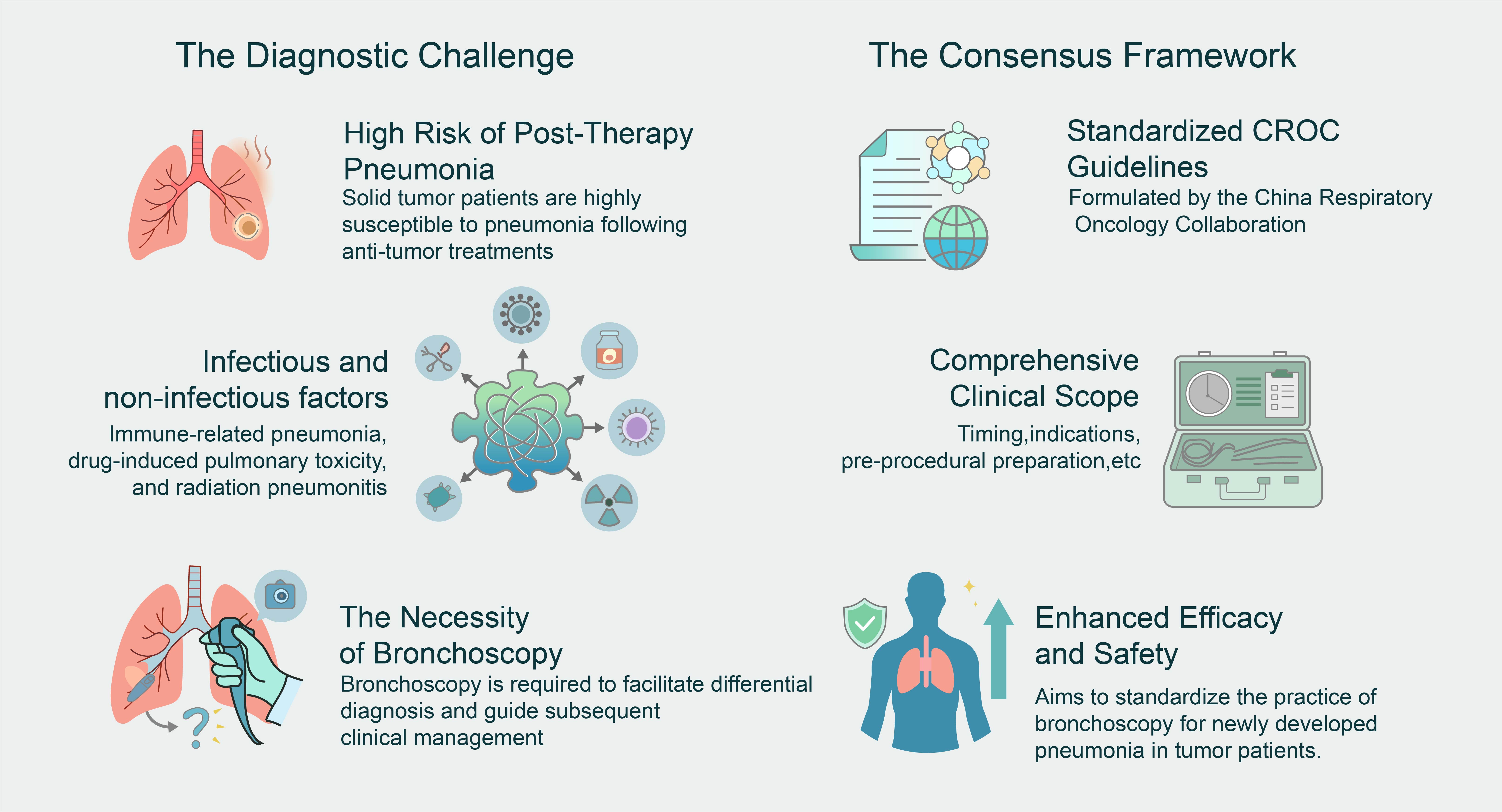
Published
Issue
Section
License
Copyright (c) 2025 Xi Chen, Jing Zheng, Jingjing Qu, Cuihong Cai, Qingyong Chen, Xueqin Chen, Zhuo Cao, Qi Dai, Yongmin Ding, Yong Fang, Dan Liu, Shaohua Lu, Renyong Guo, Hui Huang, Chenghua Jin, Mei Kong, Hui Guo, Jinpeng Liu, Min Li, Xiaodong Lv, Weiqiang Mo, Xin Su, Yilan Sun, Xiaoyu Wu, Qing Yang, Xin Yao, Jian Ye, Junhui Ye, Xiaoqun Ye, Wanjun Yu, Xianghua Ye, Hong Shen, Jingchen Zhang, Jingfeng Zhang, Xiaochen Zhang, Chenzhi Zhou, Dan Zhu, Junfei Zhu, Hua Zhong, Runbo Zhong, Zhengfei zhu, Zhen Li, Jianya Zhou (Author)

This work is licensed under a Creative Commons Attribution-NonCommercial 4.0 International License.